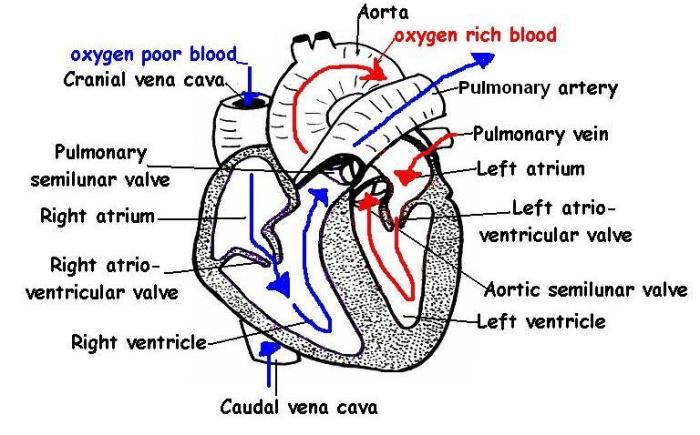
Calling all cat enthusiasts and aspiring veterinarians! The cat blood vessels labeling quiz is your passport to unraveling the intricate network that sustains feline life. Prepare to embark on a captivating journey where you’ll decipher the structure, function, and clinical significance of blood vessels in our beloved furry friends.
Dive into a comprehensive overview of feline vascular anatomy, unraveling the intricacies of arteries, veins, and capillaries. Test your knowledge with an engaging quiz that challenges your understanding of blood vessel labeling, complete with illustrative images and a handy answer key.
Anatomical Overview
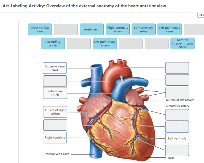
The vascular system in cats is a complex network of blood vessels that transport blood throughout the body. It plays a crucial role in delivering oxygen, nutrients, and hormones to tissues while removing waste products.
The feline vascular system consists of three main types of blood vessels: arteries, veins, and capillaries. Each type has a distinct structure and function.
Arteries
Arteries are blood vessels that carry oxygenated blood away from the heart to the rest of the body. They have thick, muscular walls that allow them to withstand the high pressure of the blood being pumped by the heart.
Veins
Veins are blood vessels that carry deoxygenated blood back to the heart. They have thinner walls than arteries and contain valves to prevent the backflow of blood.
Capillaries
Capillaries are the smallest blood vessels in the body. They form a network of tiny vessels that allow for the exchange of oxygen, nutrients, and waste products between the blood and the surrounding tissues.
If you’re looking to brush up on your cat blood vessel labeling skills, you might want to check out our quiz. We’ve got a variety of questions to test your knowledge, so you can be sure you’re ready for your next anatomy exam.
And if you’re feeling particularly ambitious, you can even try your hand at our atomic dating game answer key . Just remember, the key to success is practice, practice, practice!
Labeling Quiz
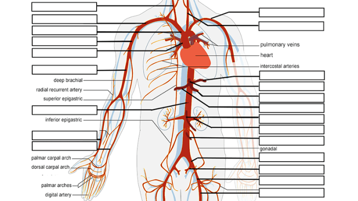
To assess your understanding of cat blood vessel labeling, engage in this comprehensive quiz.
Quiz Instructions
- Examine the provided images of cat blood vessels.
- Identify and label the indicated blood vessels.
- Submit your labeled images for evaluation.
Image Details
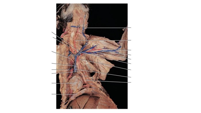
The images used in the quiz depict various anatomical regions of cats, showcasing their blood vessel networks. Each image is accompanied by a set of labels that correspond to specific blood vessels.
Answer Key
Upon completing the quiz, refer to the provided answer key or consult with an instructor to verify the accuracy of your labeling.
Histological Techniques
Histological techniques are essential for visualizing blood vessels in cats. These techniques involve the preparation and examination of thin tissue sections to study the microscopic structure of blood vessels and their surrounding tissues.
There are several histological techniques commonly used for this purpose, each with its own advantages and disadvantages:
Light Microscopy
- Advantages:
- Widely accessible and relatively inexpensive.
- Allows for the visualization of general tissue architecture and blood vessel morphology.
- Can be combined with immunohistochemistry to identify specific cell types and molecules.
- Disadvantages:
- Limited resolution compared to electron microscopy.
- Can be challenging to distinguish between different types of blood vessels.
- May require specialized staining techniques to enhance blood vessel visibility.
- Example:A light microscopy image of a cat blood vessel stained with hematoxylin and eosin (H&E) shows the general morphology of the vessel, including the endothelial cells, smooth muscle cells, and surrounding connective tissue.
Electron Microscopy
- Advantages:
- Provides ultra-high resolution, allowing for the visualization of fine structural details of blood vessels.
- Can distinguish between different types of blood vessels based on their ultrastructure.
- Can be used to study the relationship between blood vessels and surrounding cells and tissues.
- Disadvantages:
- Requires specialized equipment and expertise.
- Can be time-consuming and expensive.
- May require specialized sample preparation techniques.
- Example:An electron microscopy image of a cat blood vessel shows the endothelial cells, basement membrane, and surrounding smooth muscle cells in great detail.
Immunohistochemistry
- Advantages:
- Allows for the visualization of specific proteins and molecules within blood vessels.
- Can be used to identify and localize specific cell types and markers.
- Can provide insights into the function and regulation of blood vessels.
- Disadvantages:
- Requires specific antibodies and optimization for each target molecule.
- Can be subject to background staining and non-specific binding.
- May require specialized equipment and expertise.
- Example:An immunohistochemistry image of a cat blood vessel stained for the endothelial marker CD31 shows the distribution of endothelial cells within the vessel wall.
Clinical Applications
Blood vessel labeling in cats finds significant applications in veterinary medicine, aiding in the diagnosis and treatment of various diseases. It enables veterinarians to visualize and assess the structure and function of blood vessels, facilitating accurate diagnosis and targeted therapies.
Diagnostic Applications
Blood vessel labeling allows veterinarians to identify abnormalities in the vascular system that may indicate underlying diseases. For instance, it can help detect and diagnose conditions such as vascular malformations, aneurysms, and occlusions. By assessing the size, shape, and distribution of blood vessels, veterinarians can determine if they are functioning normally or if there are any blockages or irregularities that could lead to health issues.
Therapeutic Applications
In addition to diagnosis, blood vessel labeling can also guide therapeutic interventions. By precisely targeting specific blood vessels, veterinarians can deliver drugs or therapies directly to the affected areas. This targeted approach minimizes systemic side effects and enhances the efficacy of treatment.
For example, blood vessel labeling can be used to deliver chemotherapy drugs to tumors or to treat vascular diseases such as atherosclerosis.
Case Studies, Cat blood vessels labeling quiz
Numerous case studies have demonstrated the successful use of blood vessel labeling in clinical practice. One study reported the use of a fluorescent dye to label blood vessels in a cat with a suspected vascular malformation. The dye allowed the veterinarian to visualize the abnormal blood vessels and determine their extent, enabling a precise surgical intervention to correct the malformation.
In another case, blood vessel labeling was employed to guide the delivery of chemotherapy drugs to a tumor in a cat. The labeled blood vessels facilitated the targeted delivery of the drugs directly to the tumor site, reducing the risk of side effects and improving the chances of successful treatment.
Essential FAQs: Cat Blood Vessels Labeling Quiz
What are the key differences between arteries, veins, and capillaries?
Arteries carry oxygenated blood away from the heart, while veins return deoxygenated blood to the heart. Capillaries are the smallest blood vessels, allowing for the exchange of oxygen and nutrients between the blood and surrounding tissues.
How can blood vessel labeling aid in diagnosing feline diseases?
Blood vessel labeling techniques can visualize abnormalities in blood flow, helping diagnose conditions such as heart disease, cancer, and vascular malformations.
What histological techniques are commonly used to study cat blood vessels?
Histological techniques like immunohistochemistry and lectin histochemistry enable researchers to visualize specific proteins and structures within blood vessels, providing insights into their function and development.
